You just walked out of a sonography center, envelope in hand. Before your follow-up appointment with your doctor, curiosity gets the better of you. You pull out the ultrasound report, scan the page, and immediately freeze at the sight of words like "Hepatomegaly," "Echogenic," or "Renal Calculi."
Cue the frantic Google searching.
If you live in Mumbai's fast-paced western suburbs—from Andheri and Jogeshwari to Goregaon and Malad—you likely don't have hours to spend decoding complex medical jargon. Medical imaging reports are written by Radiologists specifically for other doctors, which is why they sound so intimidating.
At Biocheck Diagnostics in Goregaon West, we believe in empowering our patients. You shouldn't have to panic over a standard medical term. In this ultimate guide, our expert radiology team translates the most common (and most misunderstood) sonography terms into plain English.
Due to the modern Mumbai lifestyle—characterized by late-night meals, highly processed foods, and high stress—the liver and gallbladder are two of the most frequently scanned organs during a Whole Abdomen Ultrasound.
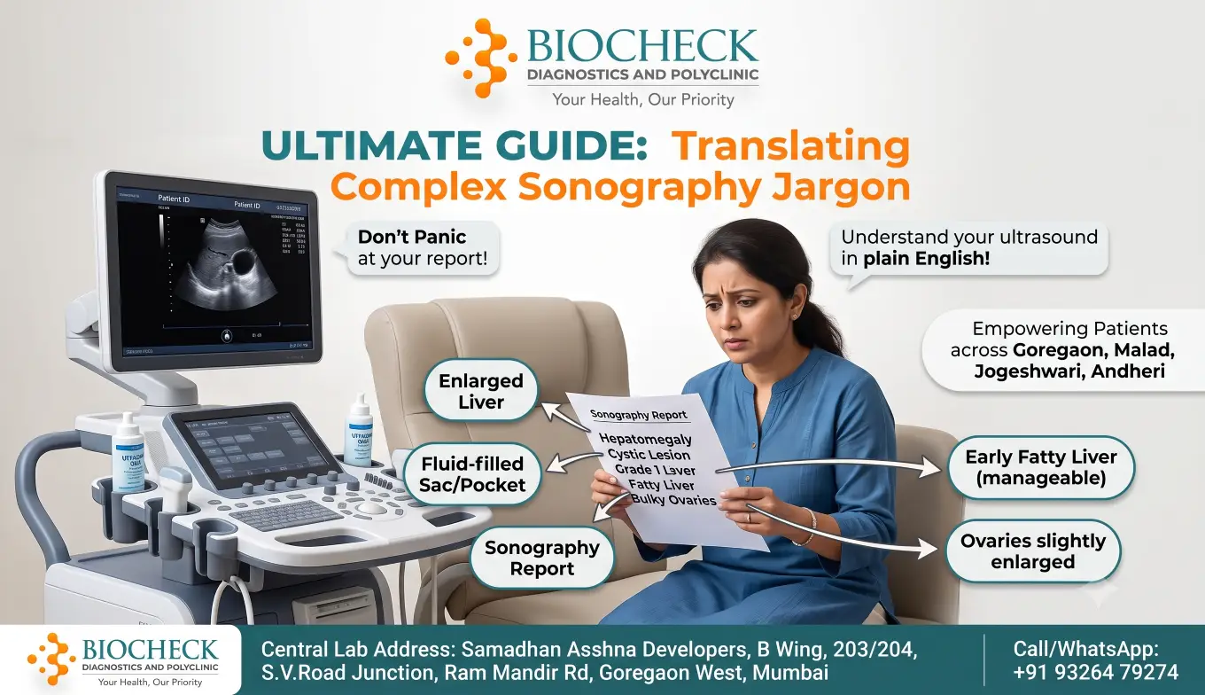
- Hepatomegaly: This simply means an "enlarged liver." It indicates that your liver is swollen beyond its normal size. While it sounds scary, it is often a reversible condition caused by diet or mild infections.
- Grade 1 / Grade 2 Fatty Liver (Hepatic Steatosis): This is incredibly common. It means there is an accumulation of fat cells inside your liver.
- Grade 1 is mild and usually completely reversible with a healthier diet and exercise.
- Grade 2 or 3 means the fat buildup is more severe and requires immediate lifestyle intervention to prevent inflammation. (Pro Tip: If you have a fatty liver, your doctor will likely recommend a Liver Function Test (LFT) and a Lipid Profile blood test to check your cholesterol. You can book these at our Goregaon lab).
- Cholelithiasis: This is the medical term for Gallstones. It means there are hardened deposits of digestive fluid forming in your gallbladder.
- Sludge (Biliary Sludge): This refers to thickened bile in your gallbladder. It isn't quite a stone yet, but it can cause indigestion and may develop into gallstones if ignored.
Dehydration is a major issue during Mumbai's humid summers and muggy monsoons, leading to a massive spike in kidney-related ultrasound findings.
- Renal Calculi / Nephrolithiasis: Do not panic—this is simply the medical term for Kidney Stones. The report will usually specify the size in millimeters (e.g., "3mm calculus") and its exact location.
- Hydronephrosis: This means one or both of your kidneys are swollen because urine is not draining properly. This is most often caused by a kidney stone blocking the ureter (the tube connecting the kidney to the bladder).
- Cortical Cyst: A simple fluid-filled sac on the kidney. The word "cyst" causes a lot of anxiety, but simple cortical cysts are almost always benign (non-cancerous), very common as you age, and usually do not require any treatment.
For women undergoing a pelvic sonography, the terminology can be particularly stressful, especially when evaluating menstrual irregularities or fertility issues.
- Bulky Uterus: This means your uterus is slightly larger than average. It is a very common finding and is frequently caused by harmless conditions like Adenomyosis or Fibroids. It does not automatically mean you have a serious disease.
- Uterine Fibroid (Leiomyoma): A non-cancerous growth of muscle tissue in the wall of the uterus. They are incredibly common in women in their 30s and 40s. The report will note their size and location (e.g., intramural, subserosal).
- Bulky Ovaries / Polycystic Ovarian Morphology (PCOM): If your report mentions "bulky ovaries with multiple small follicles," it is describing the classic appearance of Polycystic Ovary Syndrome (PCOS). It means your ovaries contain multiple small, undeveloped egg sacs that failed to release an egg. (Note: If this appears on your scan, your gynecologist will likely request a Female Hormone Profile (FSH, LH, Prolactin, Testosterone) to confirm the diagnosis).
- Endometrial Thickness (ET): The endometrium is the lining of your uterus. The thickness fluctuates naturally throughout your menstrual cycle. Your doctor uses this measurement to track ovulation or investigate heavy bleeding.
Radiologists use specific words to describe how sound waves bounce off your organs.
- Echogenic / Hyperechoic: This means a structure appears very bright white on the ultrasound screen because it reflects a lot of sound waves. (e.g., Kidney stones and bones are highly echogenic).
- Hypoechoic: The structure appears darker grey than the surrounding tissue.
- Anechoic: This means the structure appears completely black on the screen, which usually indicates it is filled with clear fluid (like a simple cyst or a full bladder).
- Lesion: In medical terms, a lesion simply means any area of tissue that is abnormal. It is a broad term that includes harmless cysts, benign moles, and scars. It does not automatically mean cancer.
An ultrasound is heavily dependent on the skill of the person holding the probe. Unlike an X-ray or MRI, which takes a static picture, sonography is a "live" test.
If you are traveling from Andheri, Jogeshwari, or Malad, you want to ensure your scan is performed by an expert. At Biocheck Diagnostics and Polyclinic, our sonography department is spearheaded by highly experienced MD Radiologists, including Dr. Shashank J. Shinde (14+ years of expertise).
We utilize advanced, high-resolution imaging technology to ensure every measurement is precise, guaranteeing that your referring doctor receives the most accurate data possible to map out your treatment plan.
While this guide helps you understand the vocabulary of your report, never attempt to self-diagnose. Only your referring physician can combine these ultrasound images with your blood tests and physical symptoms to give you an accurate medical diagnosis.
If your doctor has prescribed a sonography, skip the crowded hospital wait times. Visit Biocheck Diagnostics in Goregaon West for a comfortable, professional experience with immediate, reliable reporting.
- 📍 Ultrasound Center: Samadhan Asshna Developers, B Wing, 203/204, S.V.Road Junction, Ram Mandir Rd, Goregaon West, Mumbai, Maharashtra 400104 (Just 2 minutes from Ram Mandir Station)
- 📞 Book Your Sonography Appointment: +91 93264 79274
- 🌐 View Our Imaging Services: Sonography
Available Near You
Whether you’re in Malad, Goregaon, Jogeshwari, Andheri or any part of Mumbai, BIOCHECK Diagnostics & Polyclinic is your trusted local centre for Sonography and comprehensive Sonography services.